
The gold-standard, minimally invasive test for diagnosing conditions of the brain's blood vessels — performed by Dr. Suresh Giragani using Digital Subtraction Angiography (DSA).

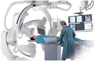
A cerebral angiogram, or Digital Subtraction Angiography (DSA), is an investigation done to delineate the structure of the brain's blood vessels and their diseases. It is performed in a dedicated angiography lab (angiosuite) equipped with DSA technology, and is widely regarded as the gold standard for diagnosing cerebral vascular conditions.
This matters most in complex cerebral aneurysms, arteriovenous malformations, dural arteriovenous fistulae, and critical stenotic or vasculitic conditions — situations where DSA remains necessary for a complete and accurate diagnosis.
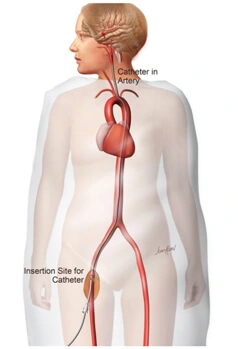
A minimally invasive investigation done through the thigh, under local anaesthesia.
A small nick is made in the groin to access the femoral artery in the thigh.
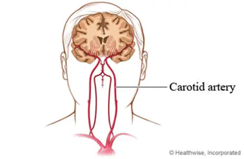
A very small plastic tube (catheter) is passed through the nick and tracked into the neck vessels.
Dye is injected into the vessel to make the blood vessels clearly visible on imaging.
Serial photographs of the brain's blood vessels are taken using DSA technology.
An empty stomach for at least 6 hours before the procedure.
Both groin regions are shaved to keep the access site clean and hairless.
Usually completed without needing overnight admission.
The full procedure is usually complete within an hour.
A cerebral angiogram, or Digital Subtraction Angiography (DSA), is a minimally invasive X-ray test that produces detailed, real-time images of the blood vessels in the brain, head, and neck. It is considered the gold standard for diagnosing cerebral vascular conditions such as aneurysms and arteriovenous malformations.
It is used to diagnose and plan treatment for brain blood vessel conditions, including aneurysms, arteriovenous malformations (AVMs), dural arteriovenous fistulae, arterial narrowing (stenosis), vasculitis, and the source of a brain haemorrhage. It is also used to guide neurointervention procedures.
The procedure usually takes 30 minutes to 1 hour and can typically be done on a day-care basis, so overnight admission is often not needed.
The procedure is performed under local anaesthesia. Only a small nick is made in the groin to access the artery, so most patients feel little more than mild pressure. There is no large incision.
Often, yes. CT and MR angiography are good screening tests but have limitations in showing fine detail and real-time blood flow. For complex aneurysms, AVMs, fistulae, and critical stenosis, a cerebral angiogram (DSA) is still needed for a complete diagnosis and accurate treatment planning.
You should have an empty stomach for at least 6 hours beforehand, and both groin areas are shaved for arterial access. Your care team will give you any additional instructions based on your health.
Patients rest and are observed for a few hours, with fluids encouraged to flush out the contrast dye. Most people resume normal activities within 24 to 48 hours, avoiding heavy lifting for a day or two.
Yes, it is considered safe when performed by an experienced interventional radiologist. It is minimally invasive with a low risk of serious complications (under 0.5%), though, as with any catheter-and-contrast procedure, a small risk exists and is discussed with each patient beforehand.
At Neuro & Vascular, cerebral angiograms are performed by Dr. Suresh Giragani, Consultant Interventional Radiologist at Apollo Hospitals, Jubilee Hills, Hyderabad, using a dedicated DSA-equipped angiography suite.
Book a consultation with Dr. Giragani, or send your scans and reports for a specialist second opinion first.